

Eye Services
Dr. Benson's practice emphasis is on treatment of glaucoma, diabetic eye dises, macular degeneration, dry eye, and other eye problems in addition to providing quality eye care to all ages.

Meet Dr. Benson
Dr. Benson is both types of eye doctors: Optometrist AND Ophthalmologist. Practicing with the motto, "Caring for you, not just your eyes," he has been attending to his patients for more than 25 years.
Frequently Asked
What is an ophthalmologist?
Why should I see an ophthalmologist?
How often should I see an ophthalmologist?
Get these answers and more.
Anatomy Of The Eye
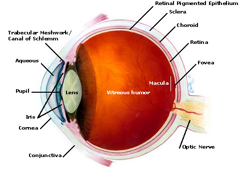
Aqueous
The clear fluid occupying the space between the cornea and the lens of the eye. The aqueous nourishes the lens and epithelial cells.
Choroid
The choroid is a layer of blood vessels that lies between the retina and sclera. These blood vessels nourish the back of the eye.
Conjunctiva
The conjunctiva is the thin, transparent tissue that covers the outer surface of the eye.
Cornea
The cornea is a clear, dome-shaped outer coating that covers the front of the eye. Light passes through the cornea to the lens. The cornea provides much of the eye’s focusing power.
Fovea
The fovea is the center area of the retina that receives the focus of an object. Nerve cells are more densely packed in this area, which allows the fovea to focus images in greater detail.
Iris
The iris is the colored part of the eye. Tiny muscles inside the iris dilate (widen) and contract (narrow) the size of the pupil, much like an aperture on a camera.
Lens
The lens focuses light onto the retina in the back of the eye.
Macula
The macula is the small and highly sensitive part of the retina responsible for detailed central vision. The macula allows us to appreciate detail and perform tasks that require central vision, such as reading.
Optic Nerve
The optic nerve transmits electrical light impulses from the retina to the brain for processing. \
Pupil
The pupil is the black, circular opening in the center of the iris. It regulates the amount of light entering the eye.
Retina
The retina is a very thin layer of light-sensitive tissues that line the inner part of the eye. It is responsible for capturing the light rays that enter the eye, converting them to light impulses and sending them to the brain for processing.
Retinal Pigmented Epithelium
A layer of pigmented cells that nourishes and supports the retina.
Sclera
The sclera is the tough, opaque tissue that serves as the eye’s protective outer layer. It is also known as the "white of the eye."
Trabecular Meshwork/Canal of Schlemm
The passageway in the eye for aqueous fluid to leave the eye.
Vitreous
The vitreous is a thick, transparent and colorless substance that fills the center of the eye behind the lens. It is composed mainly of water, comprises most of the eye’s volume and gives the eye its form and shape